Multiplex Immunofluorescence Image Analysis Services
We don’t just deliver results — we deliver the fully traceable analysis process behind them. From raw WSI to spatial biology conclusions, TerryDR is your end-to-end partner.
Explore Services →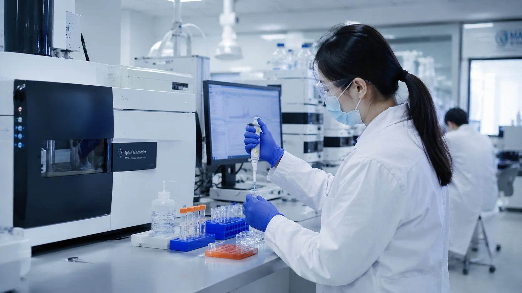
From Sample to Spatial Insight
Four flagship lines cover the full lifecycle of multiplex fluorescence pathology — analysis services, software, scanners, and stainers.
mIF Image Analysis
Traceable process data. From experimental design to data deliverables, one-stop service for multiplex fluorescence.
Learn moremIF Image Analysis Software
Nine modules cover the full spatial image analysis workflow, from preprocessing to AI-assisted exploration.
Learn moreDigital Pathology Scanner
Brightfield and multi-channel fluorescence. Full-slide digital acquisition for surgical pathology and tumor microenvironment analysis.
Learn moreAutomated IHC Stainer
Microfluidic-accelerated incubation. Uniform, consistent results in a fraction of the time.
Learn moreFrom Lab to Report, Fully Covered
Six connected stages from sample to spatially-resolved conclusions.
Sample Prep
Tissue, staining, slide prep
Staining
IHC / mIF on TR-Stainer
Scanning
5-plex on TR-sWSI
Preprocessing
TR-NoiseX · TR-StainSync
Analysis
TR-Seg · TR-PhenoClass
Reporting
Spatial heatmaps · data tables
Self-Developed Scanners, Stainers & Strips
Vertically integrated hardware — we design and build the scanners, stainers, and consumables behind every analysis.
Digital Pathology Slide Scanner
Brightfield and 5-channel fluorescence in one instrument.
3-minute brightfield scans, one-click 5-plex fluorescence configuration, Japanese optical components for stability — high value for surgical pathology and tumor microenvironment analysis.
Rapid Automated IHC Stainer
Microfluidic-chip-based. 30 minutes for full IHC, supports 40+ multiplex staining.
Reagent cost is only 1/10 of imported equivalents, supports 40+ multiplex staining.
Rapid IHC Stainer (Compact)
10 minutes instead of 3–5 hours. Compatible with standard primary & secondary antibodies.
Chip cost is 1/10 of competitors. No proprietary antibodies required.
TR-AI Assistant Wires Up 9 QuPath Modules
From image preprocessing to advanced phenotype exploration — every step in one platform.
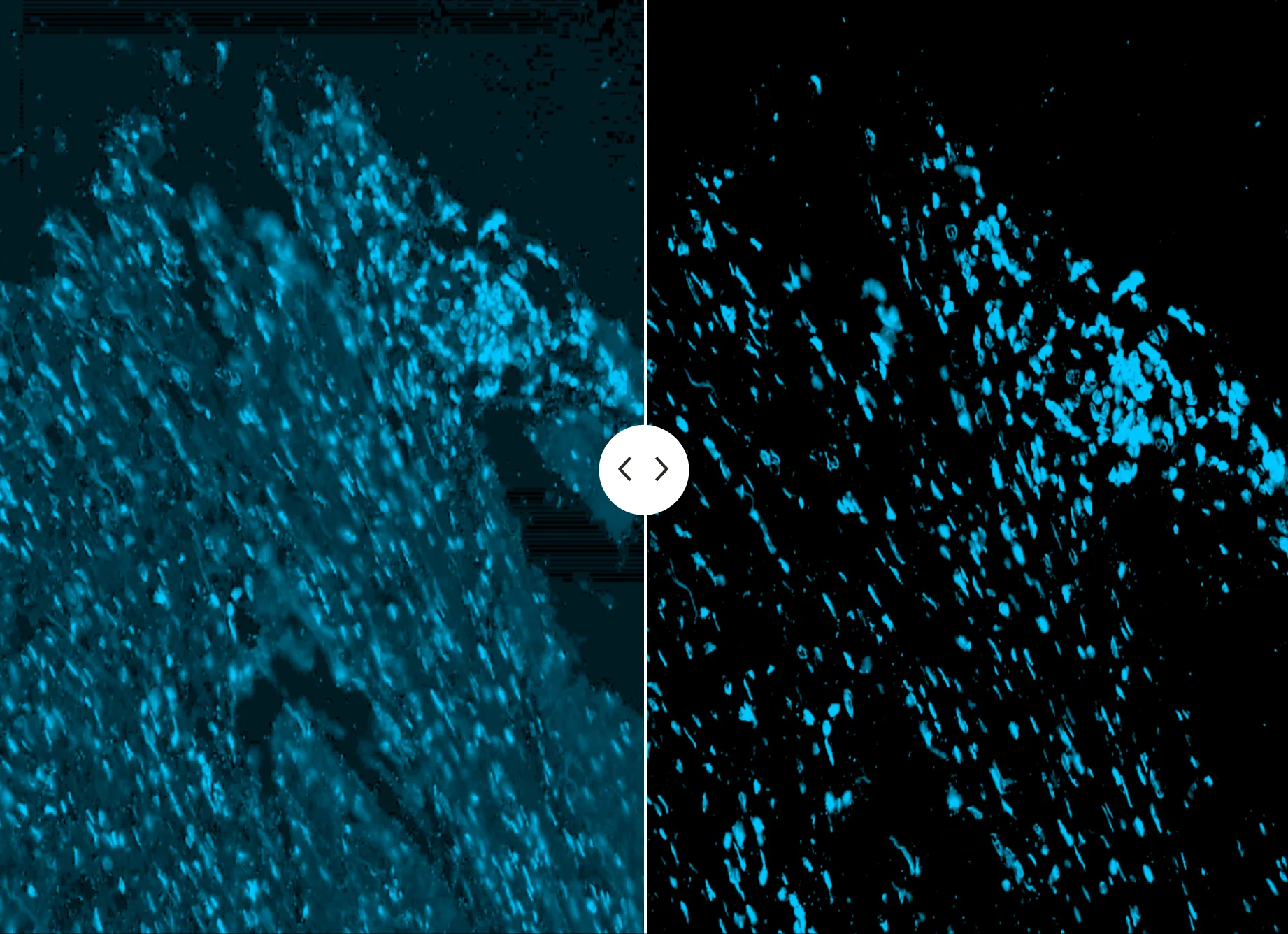
TR-NoiseX
Smart denoising, autofluorescence removal, dynamic enhancement.
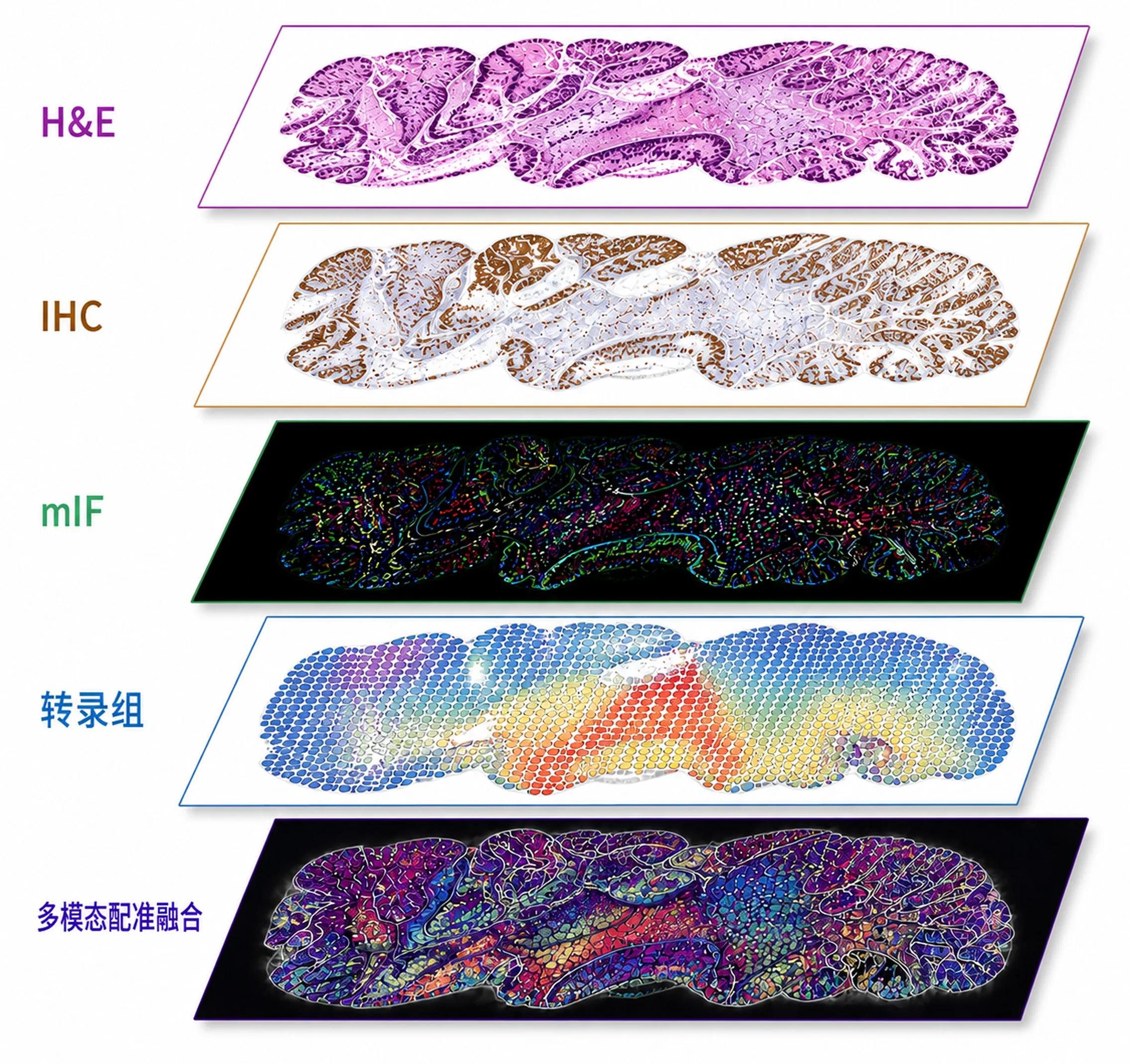
TR-StainSync
Multi-modal registration for serial sections and multi-stain images.
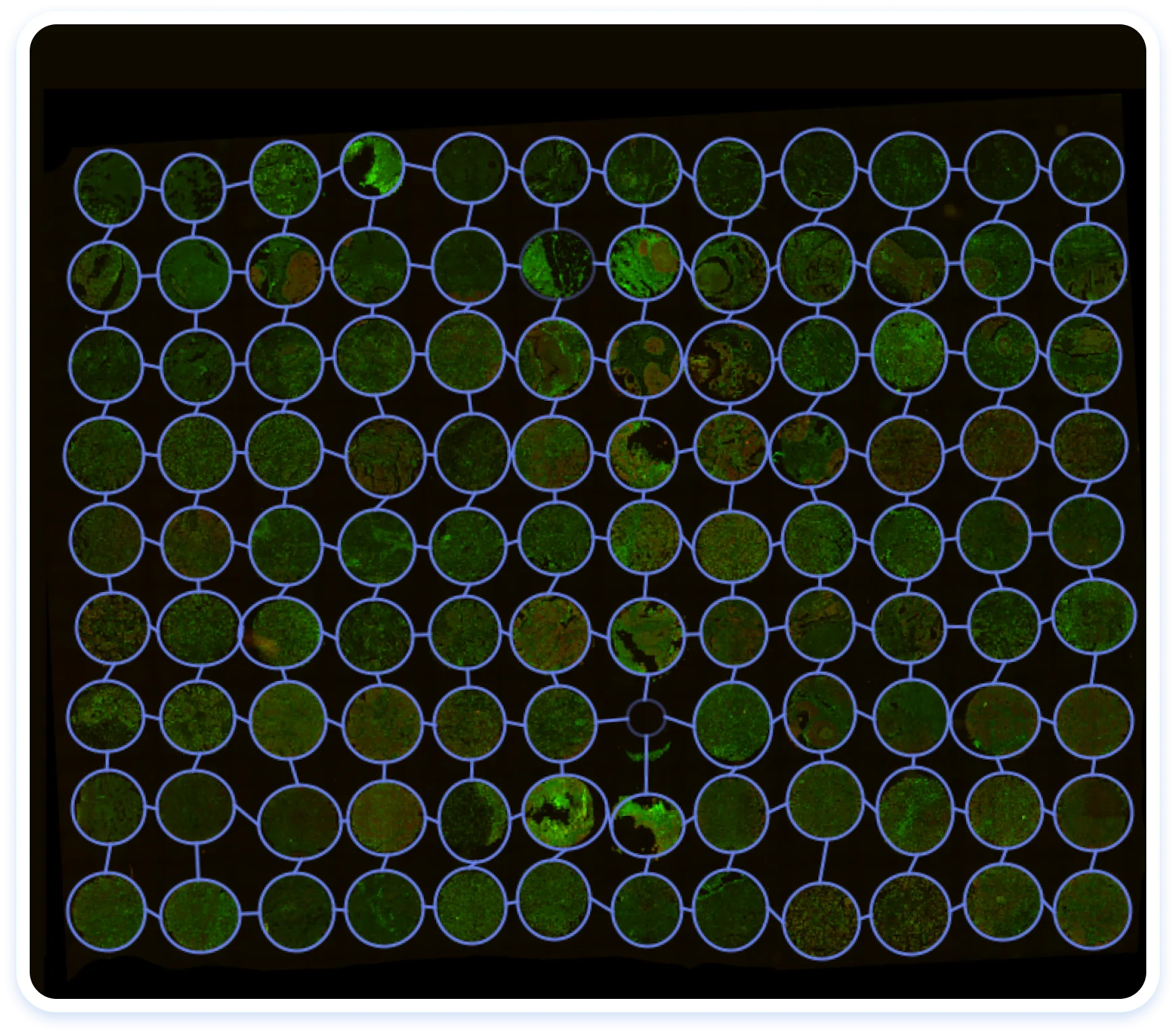
TR-TMA
Automatic tissue microarray core detection, batch processing.
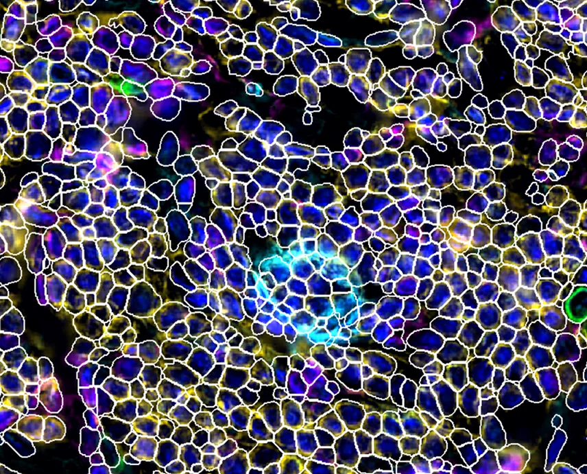
TR-Seg
Cell segmentation with nucleus, membrane, and dense-cell modes.
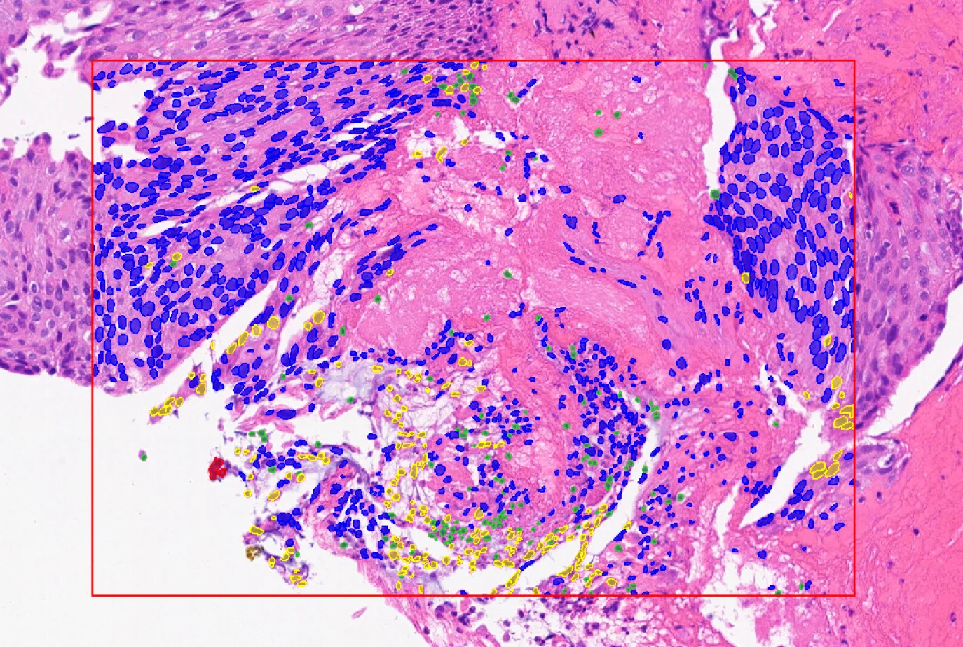
TR-HECellSage
H&E cell classification — tumor, immune, stromal, necrotic.
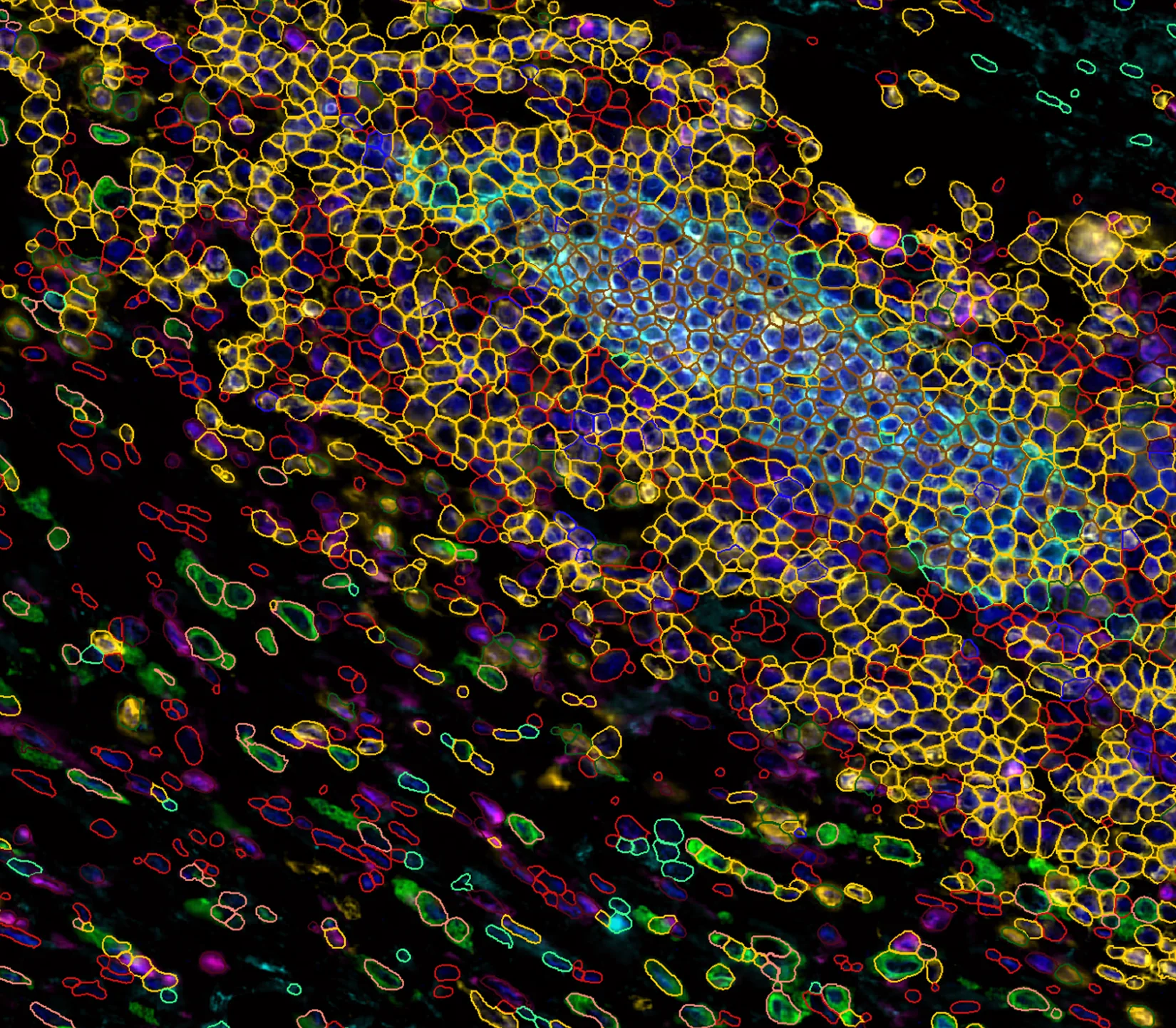
TR-PhenoClass
Deep-learning cell phenotype classification with reusable rules.
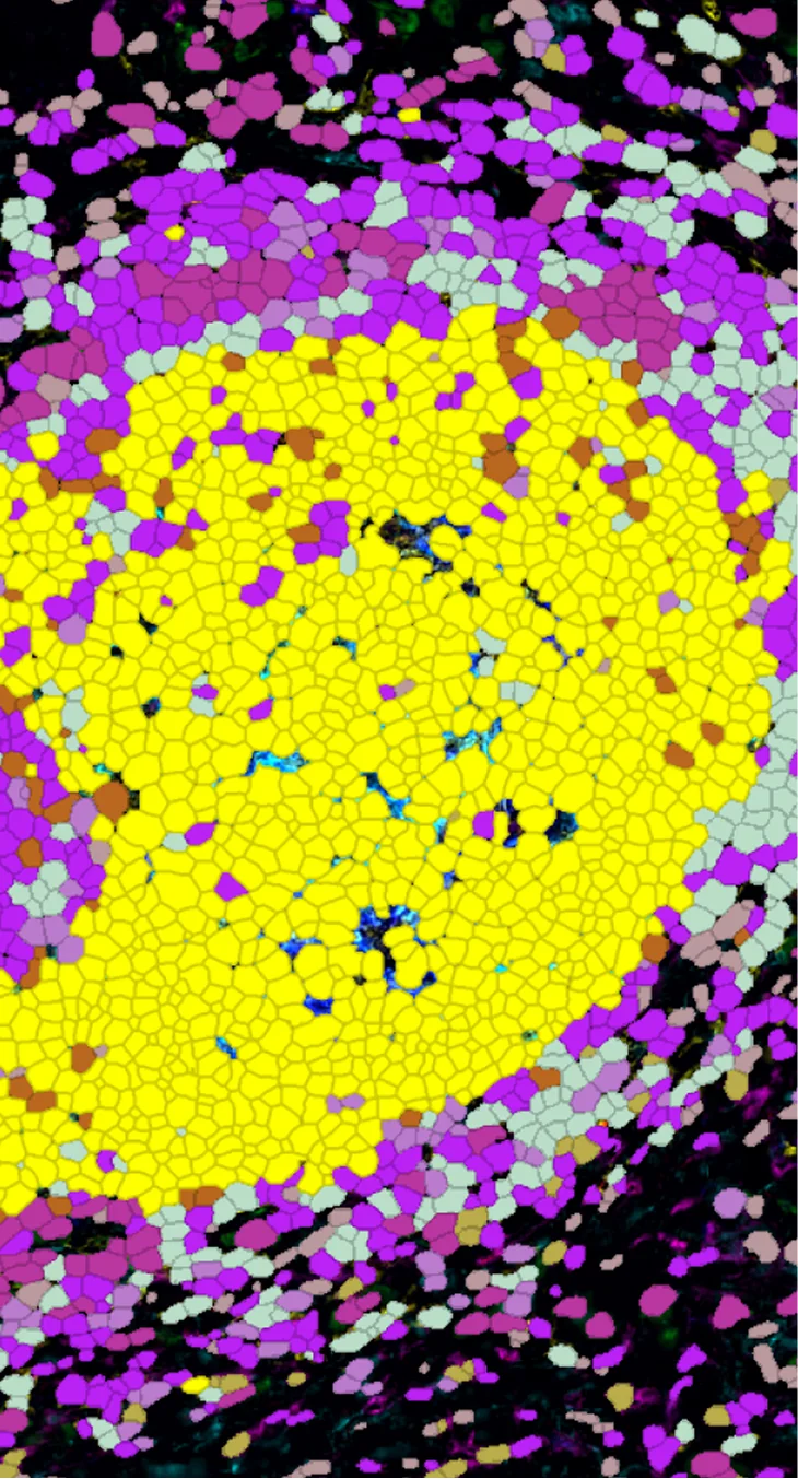
TR-PhenoDiscover
High-dimensional UMAP + Leiden clustering for rare subtypes.
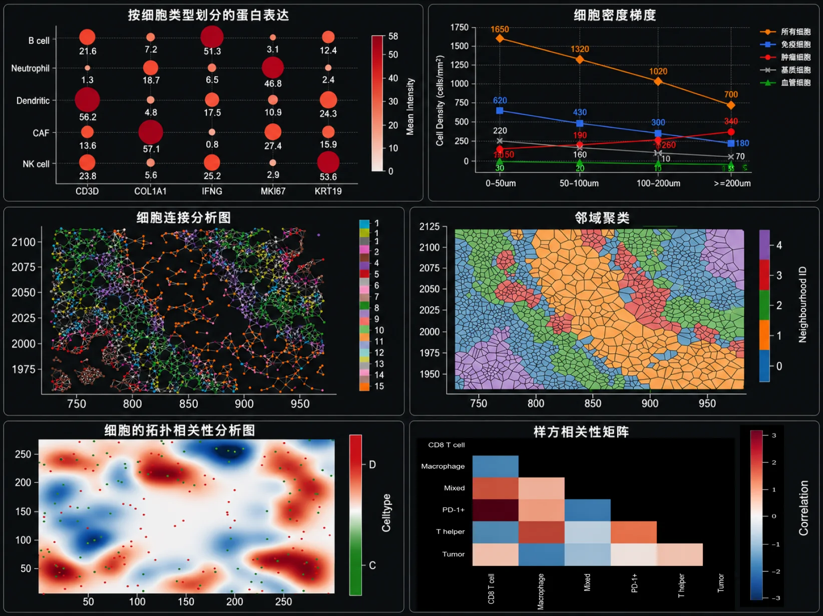
TR-Spa
Tissue spatial decoding — distances, neighborhoods, microenvironments.
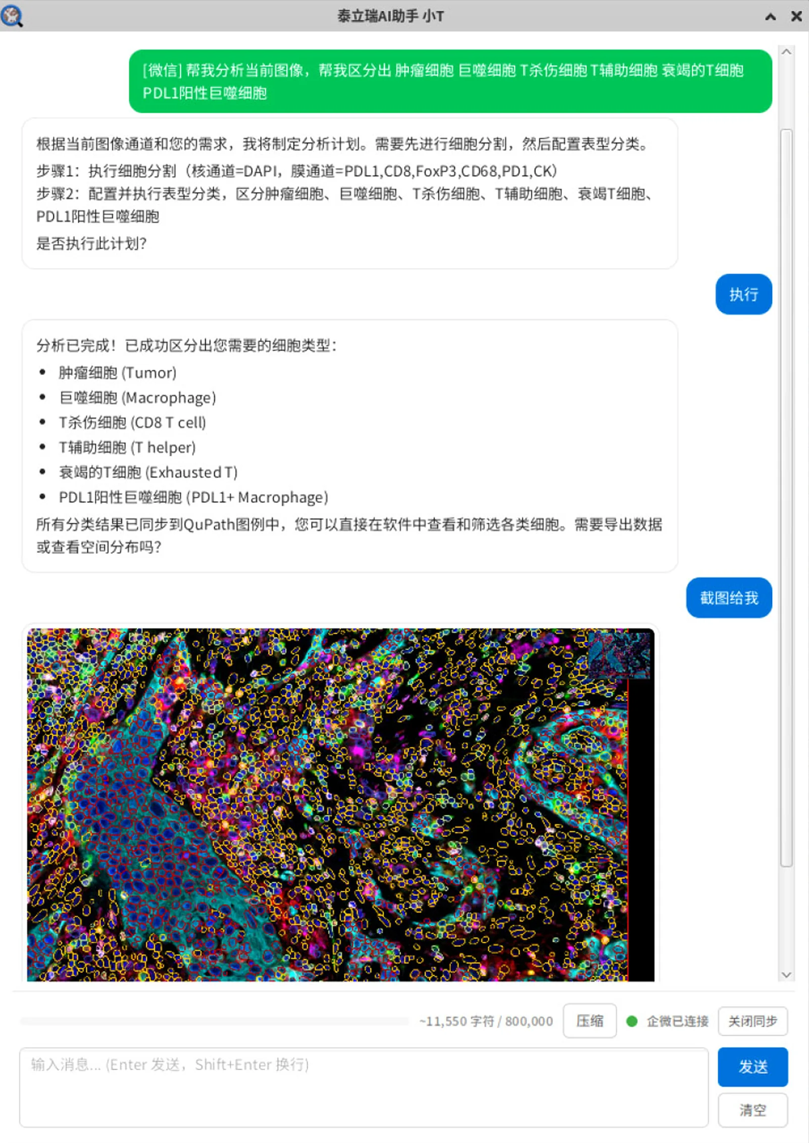
TR-AI (Assistant T)
One sentence completes the full analysis pipeline.
mIF Image Analysis
End-to-end services for digital pathology and spatial biology
- Experimental design optimization: marker design, fluorophore selection, staining order
- AI pathology analysis: registration, single-cell segmentation, phenotyping, spatial analysis
- Standardized data delivery: report generation, data matrices, chart beautification
- Algorithm customization: custom AI models, algorithm optimization, end-to-end deployment
Customized Development
OEM for devices, reagents, and spatial pathology solutions
- Device OEM: scanners, stainers, processing systems — custom development and integration
- Software OEM: spatial pathology analysis software customization and integration
Built for Hospitals, Research Labs, Pharma & More
Vertical solutions for each customer type — reagents, stainers, scanners, and software tied into a single workflow.
Hospital Pathology
Shorten stain and analysis times. Improve standardized reporting.
Learn more →Research & Universities
Support high-dimensional multi-omics data mining, accelerate spatial biology publications.
Learn more →Pharma & CRO
Reproducible analysis of batch samples, biomarker discovery and clinical translation.
Learn more →Reagent & Instrument Vendors
OEM partnerships or bundled analysis — fast validation and time-to-market.
Learn more →Third-Party Testing Labs
Standardized high-throughput workflows, stable and compliant reporting.
Learn more →Working With Leading Hospitals, Labs & Pharma
Bring us your toughest mIF sample
We’ll reply with a flat analysis plan within one business day.
