
Ultra-multiplex Spatial
Staining & Scanning System
A domestically developed, fully independent microfluidic integrated platform — a one-stop, fully automated solution from tissue section to ultra-multiplex spatial image.
 Microfluidic Cyclic Staining
 5-Channel High-Speed Scanning
 Fully Integrated Workflow
 High Throughput & Repeatability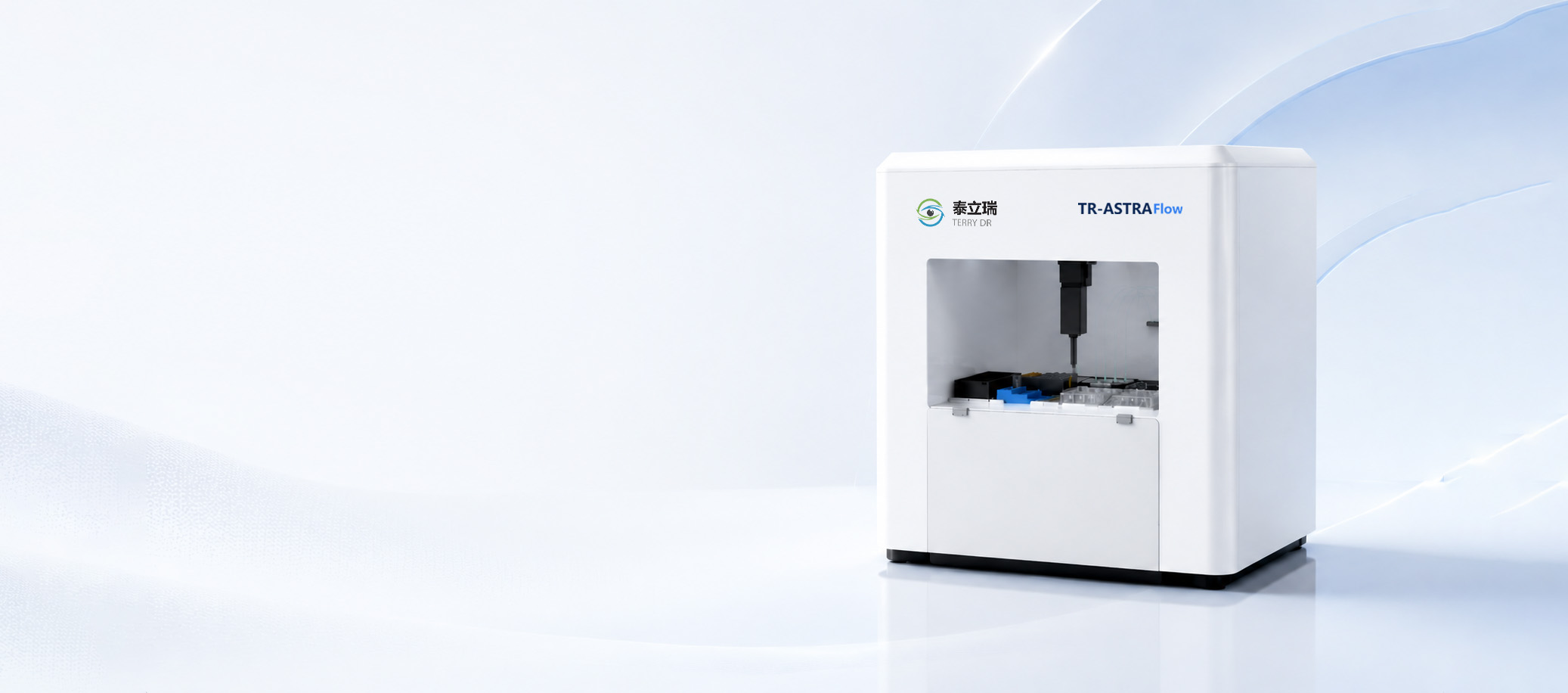

One-Stop Fully Automated Spatial Omics Workflow
Load the slide, press start, and walk away — the instrument runs the entire process unattended.
Experiment Scheme Auto-Construction
- Import antibodies or choose a standard panel
- Multiplex marker combinations generated automatically
- Covers oncology, immunology and neuroscience
Microfluidic Cyclic Staining
- Incubation, imaging and elution run automatically
- Precise control guarantees staining consistency
In-situ Whole-Slide Imaging
- Full-process scanning completed inside the instrument
- 5-channel simultaneous fluorescence imaging
- OME-TIFF standard output
Spatial Data Auto-Generation
- Automatic background correction & multi-round co-registration
- Spatial omics data exported directly
- Compatible with mainstream third-party analysis platforms
Sample Reuse Capability
- Gentle elution protects tissue architecture
- Supports multiple rounds of repeat detection
- H&E / IHC / RNA secondary analysis supported
Fully Automated, Continuous Batch Operation
One loading, and the instrument completes the entire closed loop. After antigen retrieval it switches to a fully enclosed automatic mode — no manual slide transfer at any point.
 01 Staining
 02 Imaging
 03 Elution
 04 Image Preprocessing
Calculated on a 17 × 19 mm effective area — stable fit for large-cohort spatial omics studies.
Maximum number of protein markers detected on a single section.
A mature, standardized panel library ready to deploy.
Maximum number of slides loaded in a single batch.
Staining and scanning both completed in situ — the slide never leaves the instrument.
Built for Every Research Scenario
From discovery science to regulated clinical trials — one platform covering the full translational continuum.
Basic Research
Covering neuroscience, basic immunology, infectious disease and developmental biology — map single-cell spatial phenotypes and screen entirely new molecular markers.
Translational Medicine
Spatial quantification of immune cell subsets in the tumour microenvironment and co-localization of immune checkpoint proteins — supporting targeted and immunotherapy target validation.
Clinical Sample Research
Batch multiplex staining of FFPE archival clinical samples; assess immune typing and prognostic stratification; run large-sample spatial atlases at low cost.
Clinical Trials
Spatial comparison of tissue samples before and after treatment; quantify immune cell infiltration and cell–cell interaction distance to support clinical efficacy data output.
Three Core Technology Engines
Chip, optics and software all developed in-house — the reason TR-ASTRA Flow can run staining, imaging and elution in a single enclosed loop.
BiFeX™ Microfluidic Cyclic Staining
Bidirectional microfluidic exchange chip design that cycles staining on one slide without moving it.
sWSI Five-Channel Whole-Slide Imaging
True whole-slide fluorescence acquisition with sub-pixel registration across every cycle.
Full-Process Intelligent Control
One scheduler orchestrates the whole loop and hands you analysis-ready data.
Sample & Hardware Specifications
Full technical parameters for sample handling, optics, fluidics, throughput and ecosystem compatibility.
Sample & Imaging Capacity
| Sample type | FFPE / Frozen / Single-cell suspension |
|---|---|
| Standard slide | 75 × 25 mm |
| Effective imaging area | 17 × 19 mm |
| Max slide size | 76 × 52 mm |
| Max imaging area | 34 × 19 mm |
Optical Imaging System
| Objective | 20× plan apochromat, NA 0.8 |
|---|---|
| Spatial resolution | 200 nm / pixel |
| Field of view | > 0.9 mm² |
| Fluorescence channels | DAPI / FITC / TRITC / Cy5 / Cy7 |
| Output format | OME-TIFF |
Microfluidic Circulation System
| Core technology | BiFeX™ microfluidic cyclic staining |
|---|---|
| Staining modes | Direct / Indirect / TSA |
| Incubation time | 2–8 min |
| Fluidic control | μL-level precision |
| Detection capacity | Expandable to 50-plex over multiple rounds |
Automation & Throughput
| Automated steps | Staining / imaging / elution / image preprocessing |
|---|---|
| Standard scheme | 40-plex panel system |
| Throughput | 20 slides / week |
| Operating mode | Fully enclosed, unattended, continuous batch |
| Slides per loading | Up to 4 |
Multi-omics Expansion
| Spatial proteomics | Up to 50-plex marker detection |
|---|---|
| Co-detection | RNA + protein spatial detection |
| Analysis capability | Multi-omics co-localization analysis |
| Research scenarios | Basic research / translational medicine / clinical research |
System & Ecosystem Compatibility
| In-house stack | Chip / optics / software / reagents |
|---|---|
| Antibody system | Open & compatible — no pre-conjugation required |
| Experimental standard | Standardized SOP system |
| Analysis software | QuPath / HALO / Visiopharm |
See TR-ASTRA Flow in your lab
Book a demo — send us your panel and we’ll return a run plan and sample report.