
Beyond Basic Quantification in mIF Image Analysis
 Deep Spatial Analysis
 Expert Bioinformatics
 Proprietary Software
 Isolated Client Accounts
 Process Data Delivery
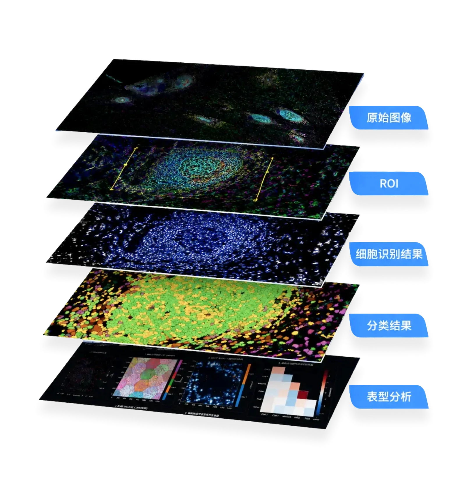
From Image to Spatial Conclusion
Full coverage of image processing, cellular phenotyping, spatial analysis, neighborhood interactions, microenvironment typing, and clinical linkage.
Multiplex Fluorescence — The Low-Hanging Fruit of High-Quality Papers

Multi-Dimensional, More Complete
Capture dozens of markers simultaneously. Reveal cellular identity, not just intensity.

Spatial Relationships, Deeper
Understand how cell types interact, where they cluster, and how neighborhoods differ.

Mechanistic Insight, Stronger
Move beyond simple counts. Link phenotypes to clinical outcomes with quantified evidence.
Eight Projects We Routinely Support
From tumor microenvironment to drug discovery — our analysis pipelines apply wherever spatial biology matters.
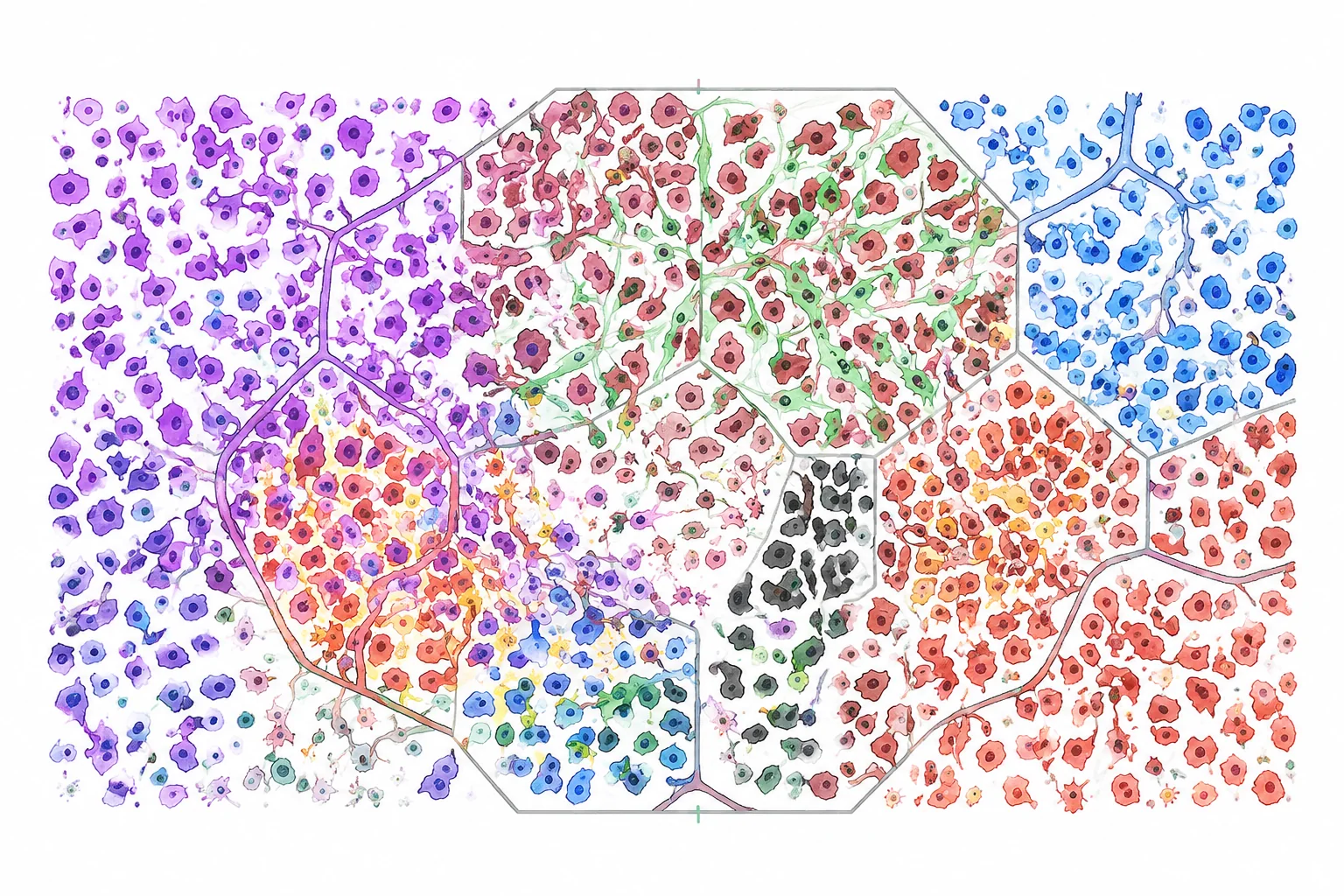 01
01Tumor Microenvironment Profiling
Quantify tumor, immune, stromal, and vascular cell composition and spatial distribution.
 02
02Immune Phenotype & Functional State
Identify multi-marker cell phenotypes — activation, cytotoxicity, proliferation, exhaustion.
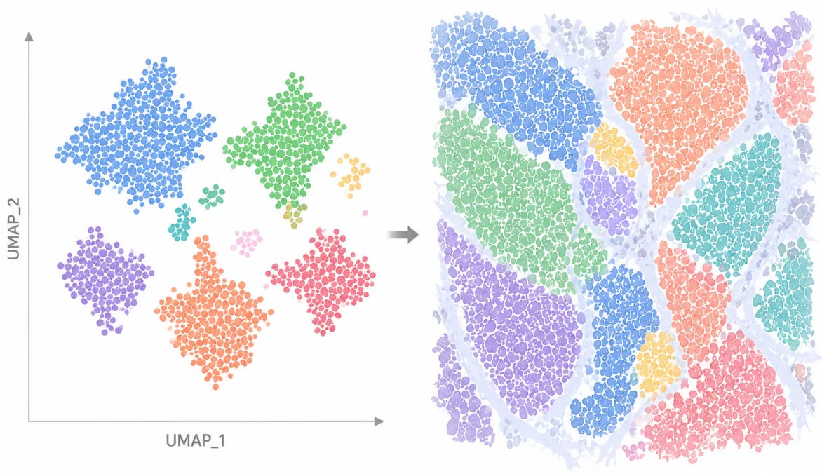 03
03Tumor Heterogeneity & Subtypes
Combine phenotyping, UMAP, and clustering to discover regional differences and rare subsets.
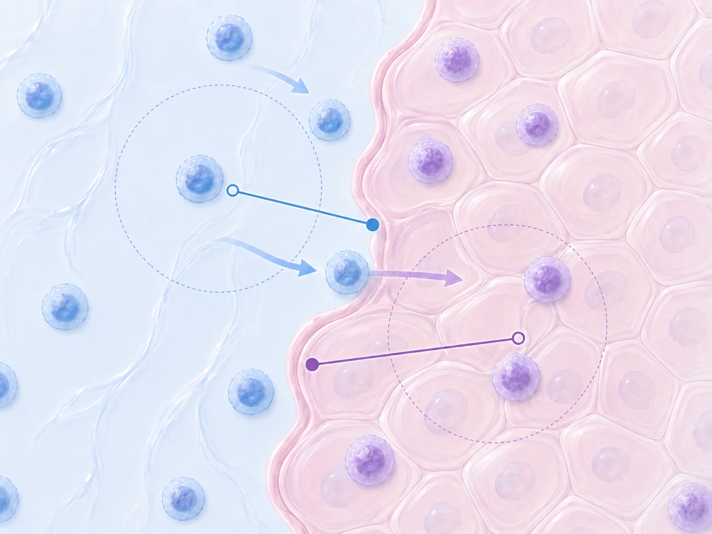 04
04Immune Infiltration & Spatial Interaction
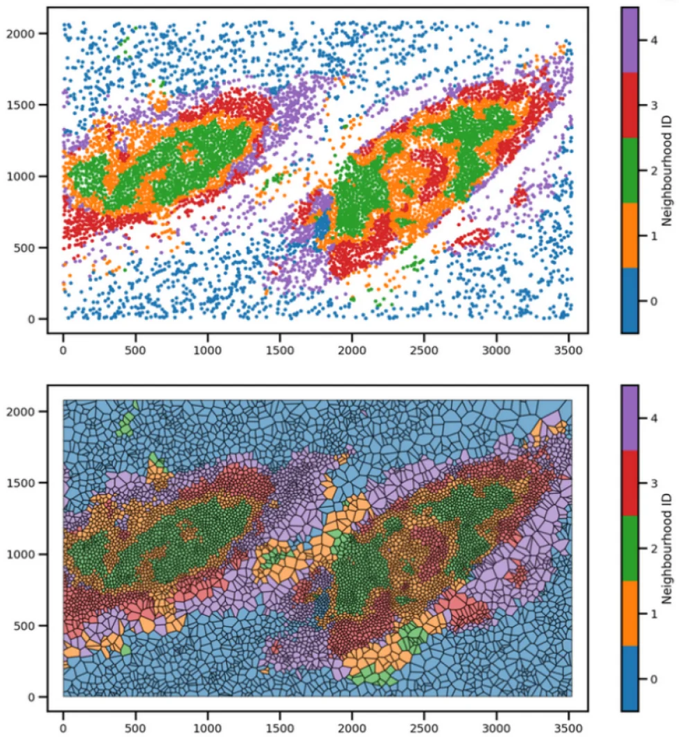
Quantify infiltration, distances, attraction/repulsion, and local neighborhood relationships.
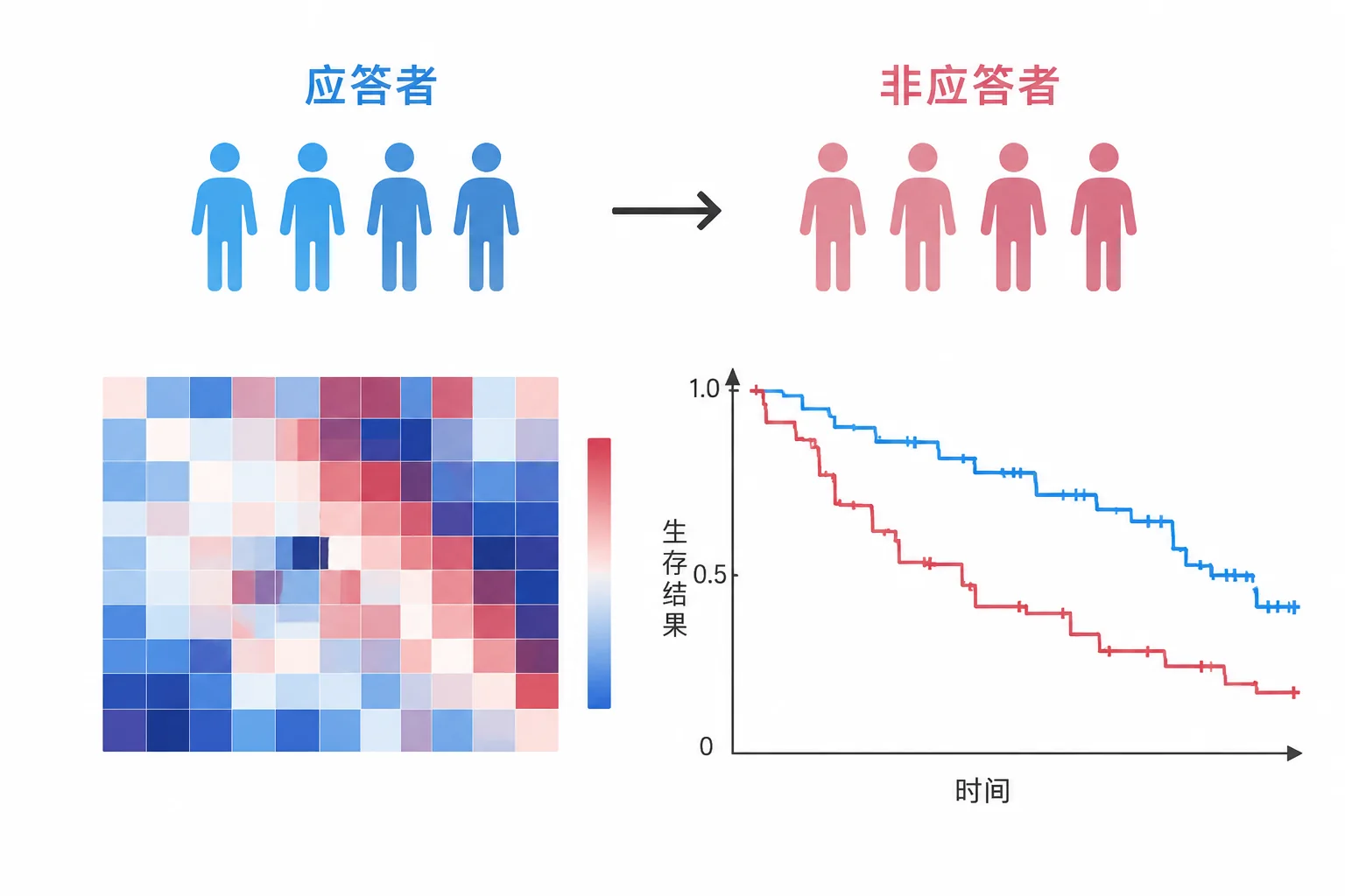 05
05Treatment Response & Biomarker Research
Compare efficacy groups, explore response, and identify prognostic spatial signatures.
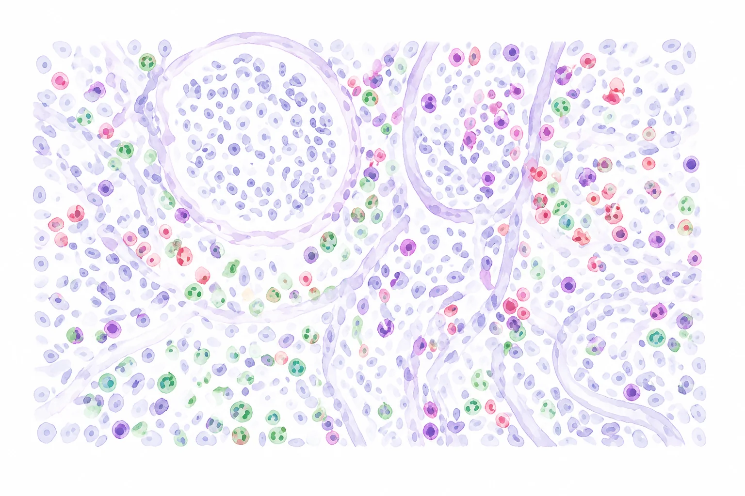 06
06Inflammation & Organ Disease
Immune infiltration, lesion structure, tissue damage, and fibrosis processes.
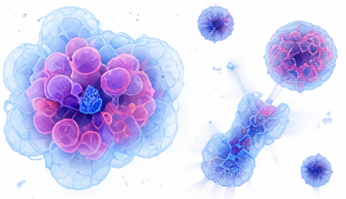 07
07Development, Organoids & Regeneration
Cell differentiation, spatial structure, tissue repair, and transplantation integration.
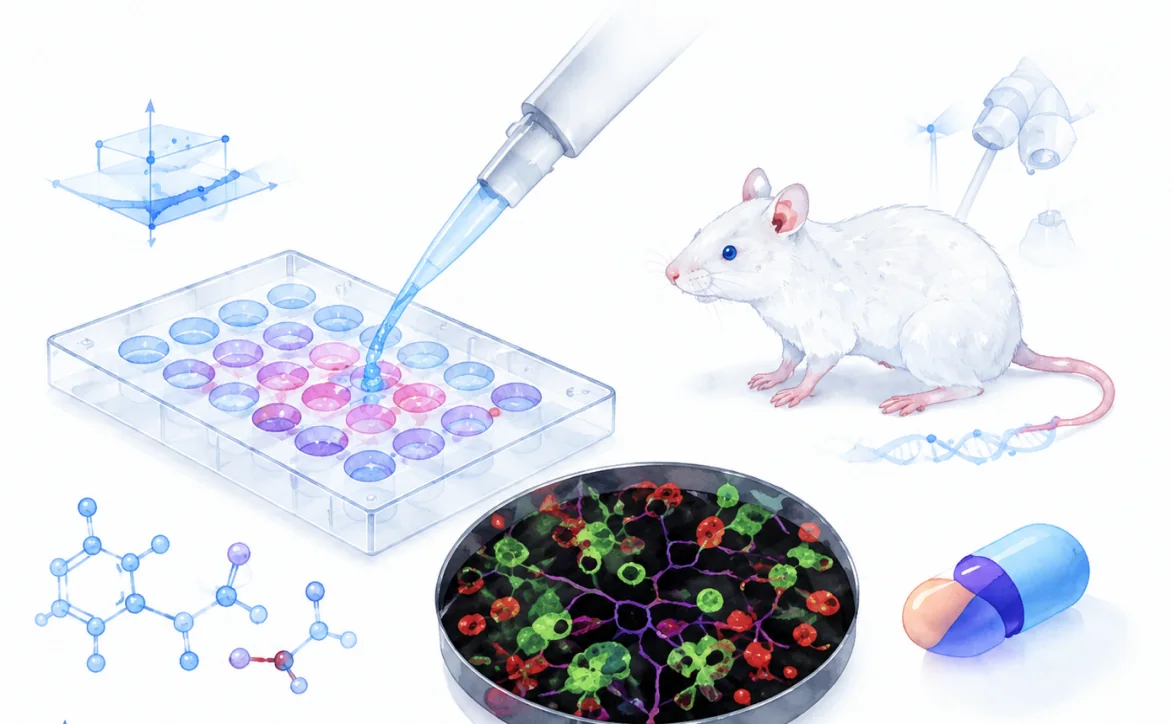 08
08Drug Development & Pre-Clinical Evaluation
Target validation, efficacy analysis, animal models, and tissue toxicity studies.
Validated in Peer-Reviewed Science
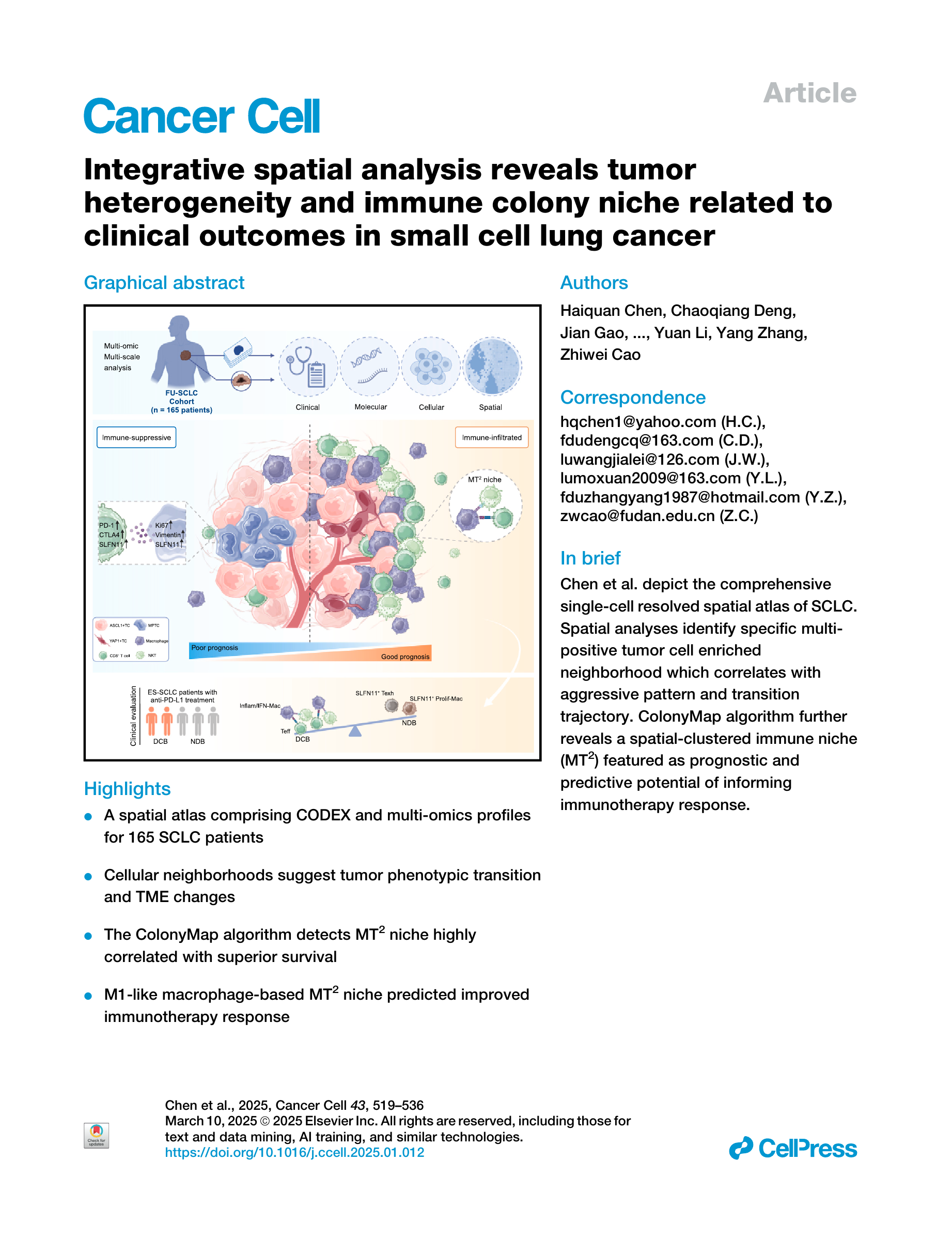
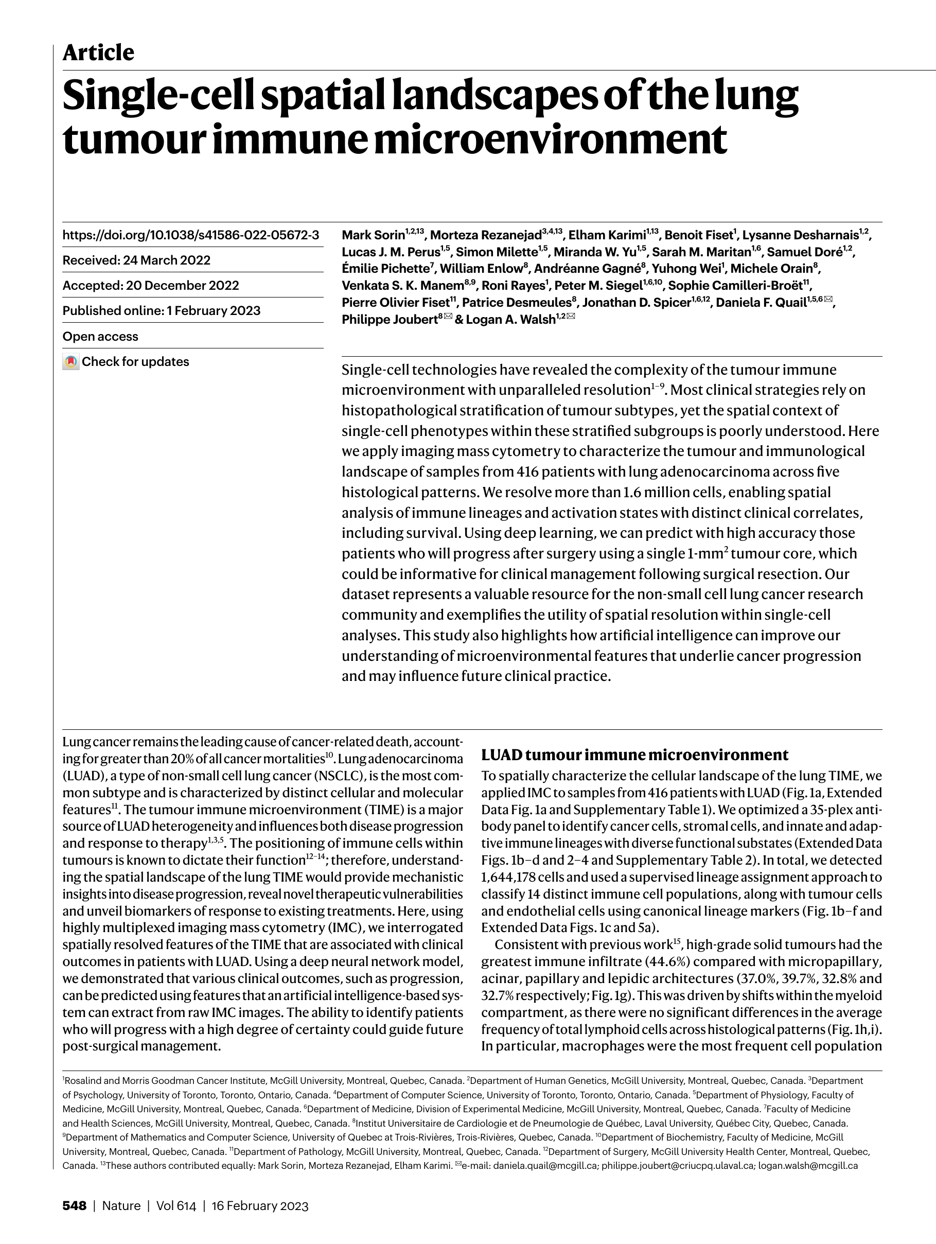
Our methods build on and contribute to the spatial biology literature — from tumor microenvironment quantification to integrative single-cell spatial analysis.

Problems We Solve, One by One
Five concrete pain points — and how TerryDR’s team and process address each.
Your Challenge
TerryDR’s Answer
Bioinformatics + Algorithm Development Under One Roof
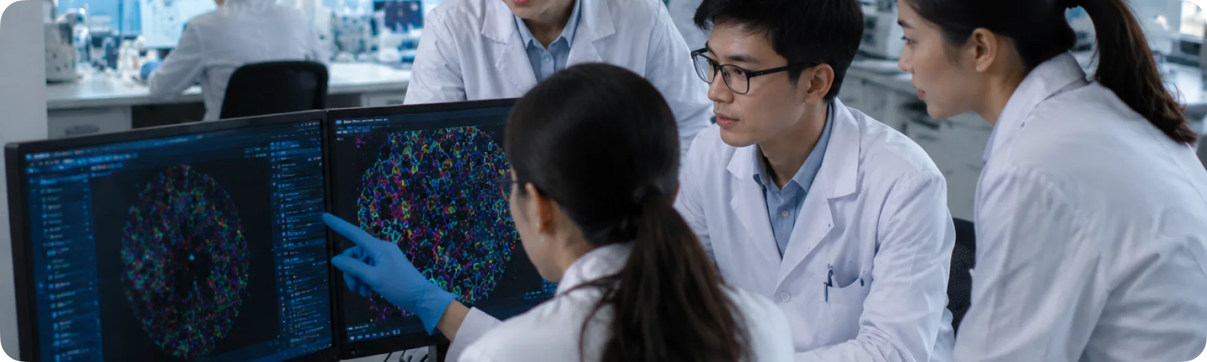
Bioinformatics & Image Analysis Team
- Stays current on multiplex fluorescence, spatial biology, and digital pathology methods
- Designs analyses around your research question, not just generic pipelines
- Hypothesis-driven + data-driven mining to fully extract value from your images

Algorithm & Software Development Team
- Complex image processing, cell detection, spatial relationship analysis
- Custom development tailored to project requirements
- Not just software users — we optimize the algorithms themselves
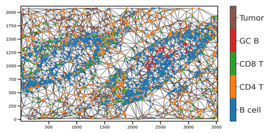
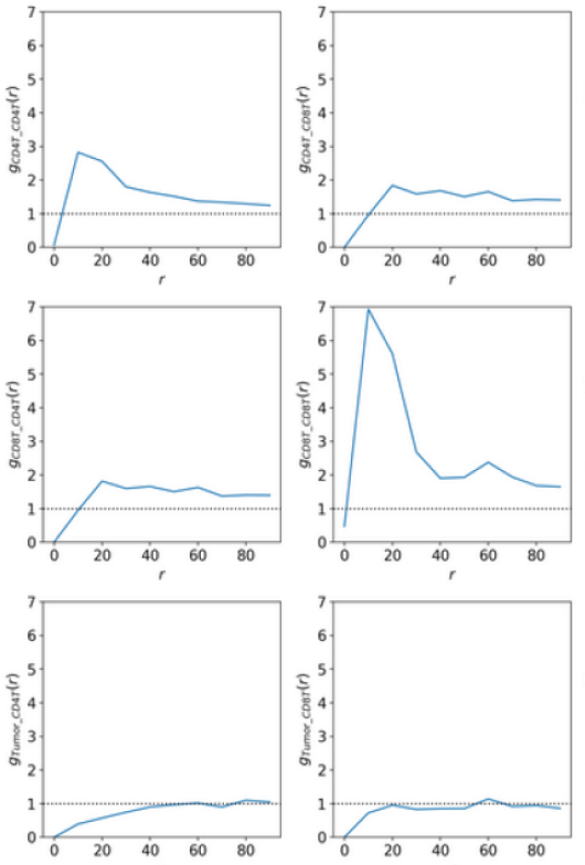
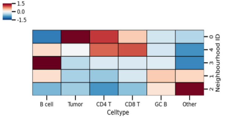
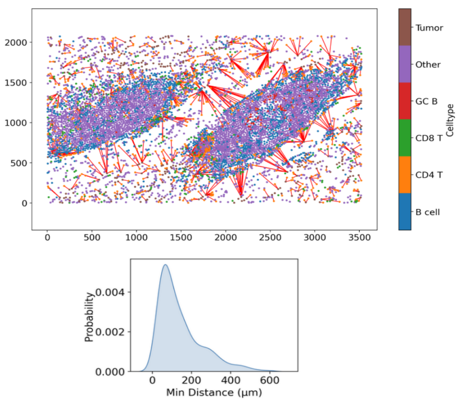
See the Analysis Pipeline at Work
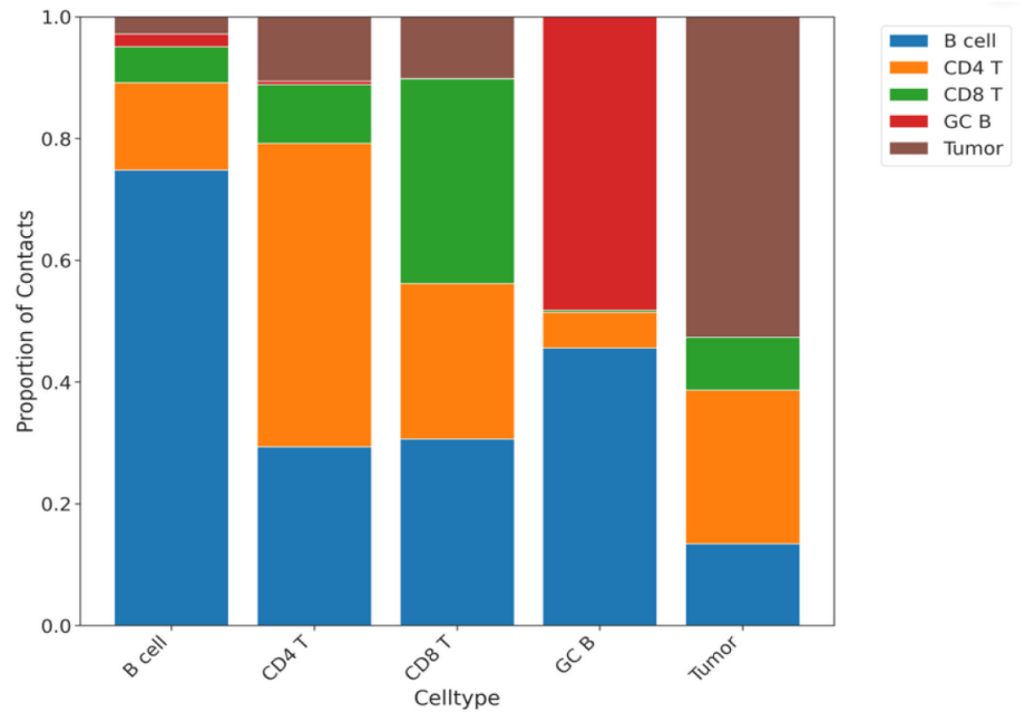
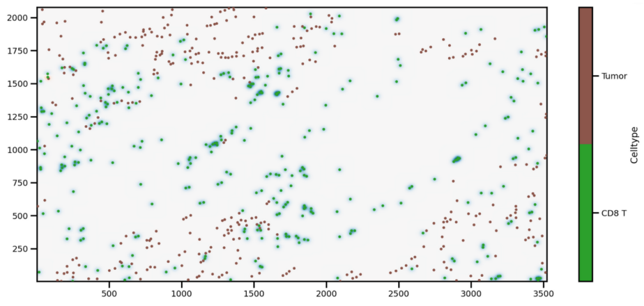
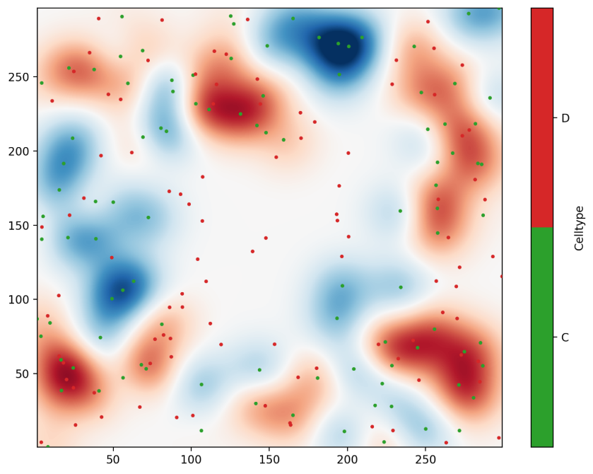
From whole-slide import to phenotype maps and spatial heatmaps — every step is visualized and exportable.
Not Just Results — Results + Process + Evidence
Traditional services only deliver “results.” TerryDR delivers the full evidence chain.
Results Cross-Verified with Images
Every quantitative result is validated against the raw image, ROI, and detection outputs.
Isolated Client Accounts
Each client has a separate project account. Permissions are clean. Data never co-mingles.
Cloud Retention & Re-Verification
Cloud storage for long-term retention, on-demand audit and traceability.
Every Step is Controllable, Every Action is Documented
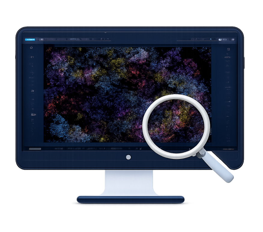
Project Discussion
Sample type, staining, image format, research goal, expected metrics.

Image Assessment
Image quality, channel configuration, tissue structure, analysis feasibility.
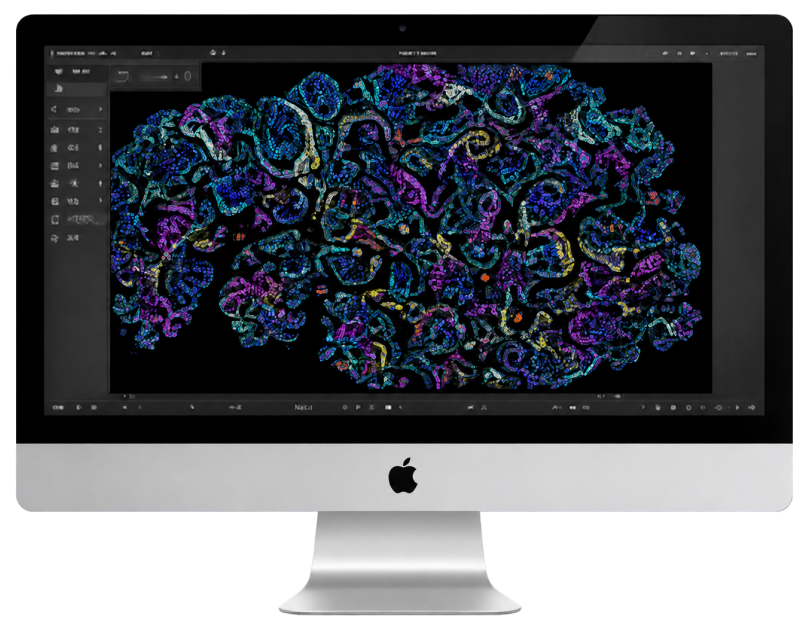
Pipeline Design
Quantitative metrics, spatial methods, output formats — all aligned to your goal.

Image Analysis
Cell detection, region segmentation, metric extraction, spatial analysis.

Process Data Delivery
QuPath cloud server delivers raw images, ROIs, detection results, and intermediate data.

Result Delivery & Interpretation
Final tables, visualizations, and analysis report — with audit and discussion support.
Make every mIF image worth more
Tell us about your study — we’ll respond with a flat analysis plan within one business day.